


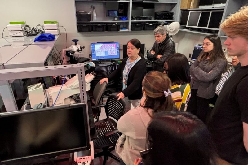
Dr. Sabine Mai leads a tour through the Nano and Cell Imaging Facility’s new space during its grand opening.
Nano and Cell Imaging Facility now on Bannatyne campus
The Rady Faculty of Health Sciences’ Nano and Cell Imaging Facility (NCIF) has a new home on the Bannatyne campus.
After three years of planning and renovations, the facility has moved from CancerCare Manitoba’s building to the department of physiology and pathophysiology’s area on the fourth floor of the Basic Medical Sciences Building.
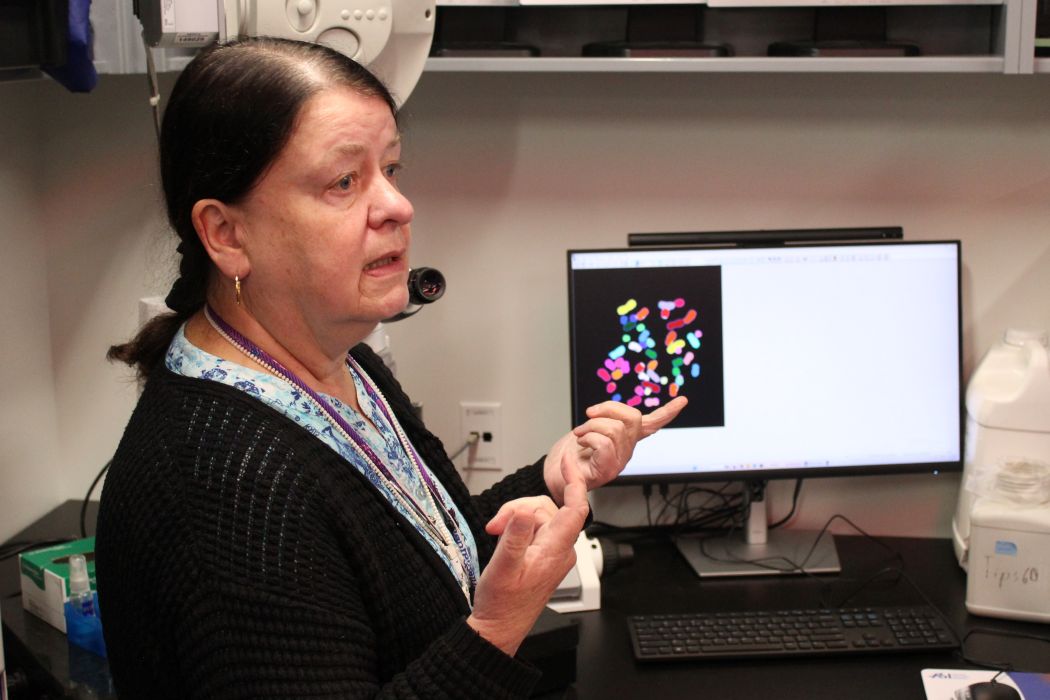
Dr. Sabine Mai, director of the Nano and Cell Imaging Facility, said it’s wonderful to have a new space on campus with cutting-edge imaging technology.
Dr. Sabine Mai (centre) speaks about the Nano and Cell Imaging Facility’s new space on the fourth floor of the Basic Medical Sciences Building.
“We have three rooms that have been completely renovated to make space for the facility. We redesigned the space to house the equipment,” said Mai, a professor of physiology and pathophysiology in the Max Rady College of Medicine, Rady Faculty of Health Sciences, and UM Canada Research Chair in genomic instability and nuclear architecture in cancer.
A grand opening took place on Oct. 15 with presentations, tours and hands-on experiences.
The facility is available for students, faculty and researchers from across Canada and around the world to use.
The newest piece of equipment is a ZEISS Elyra 7 super-resolution microscope, which has a resolution below 20 nanometers. Mai said this allows the user to see a single molecule.
Modern microscopes aren’t only for viewing specimens, she said, but also include software and cameras.
“You don’t only see – you document what you see. And that’s where the camera and software come in,” Mai said.
The facility also offers 3D imaging, laser microdissection, spectral karyotyping and advanced live cell imaging.
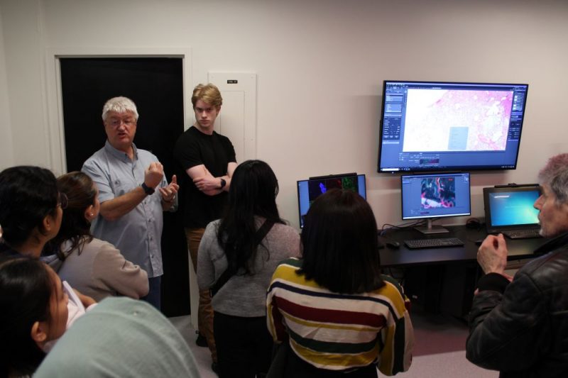
Darryl Dyck, NCIF’s expert imaging technician, speaks about the equipment in the Nano and Cell Imaging Facility.
In addition to providing researchers with the use of microscopy equipment and training opportunities, NCIF experts can help scientists design their experiments and integrate imaging into their work in any area of research.
“It can be cell biology. It can be any disease where you need to look at tissues or at individual cells. It can be a mouse model that you study. It doesn’t really matter what the object that you study is, as long as you have cells and tissues that you want to investigate,” Mai said.
The facility was opened by Mai 26 years ago to meet the growing demand for high-end microscopy. It has continuously evolved, incorporating new and cutting-edge technology.
“I was hired by the University of Manitoba because of the work that I had been doing back in Europe. I came with a single microscope, and then everybody wanted to use it, so I thought, ‘OK, if I cannot do my work because everybody’s using it, either we find another way, or I have to go back to Europe,’” Mai said.
“It was my dream in 1999 to create a facility like this. We are now one of the longest-established imaging facilities in Canada.”
Mai said that people shouldn’t be shy and should reach out to her if they are interested in learning more and want to take advantage of the imaging possibilities.
“If you have an idea about what you would like to do related to any type of imaging, just contact me,” said Mai, who can be reached at sabine.mai@umanitoba.ca.