I look prettier from my right side
Seeing mysteries and wonders of the natural world
The true mystery of the world, Oscar Wilde said, lies in the visible, not the invisible. What does shark skin look like; or pollen, a proboscis, a leaf, your lunch, a virus, the underside of a thistle leaf? Shark scales, one professor says, look like birds in flight. The underside of a thistle leaf: Like a jungle of choking vines. To see this reality though, one must look extremely close.
Daily, in a quiet lab on the fifth floor of Buller, the visible mysteries of life are revealed in the Biological Imaging Facility. Designed with the scientist in mind, it provides technologies for stimulating creativity and advancing innovative research in numerous scientific realms. Besides the user-friendly layout, the equipment is spectacular; the latest piece of kit, for example, allows a user to focus in on an individual cell in a sample and direct a laser to cut out that one cell, dropping it in a collecting tube below. The lab is a core facility of the department of biological sciences with members of the department of microbiology participating in it. The facility invites firms or researchers concerned with biomedical, biodiagnostic, or biological research to use its services.
UM Today will visit this laboratory’s work in greater depth in 2014, but for now, biological sciences professor Erwin Huebner and lab technician Andre Dufresne have selected these images to share, if for no other reason, than to inspire us to focus in on mysteries and wonder.
Mosquito
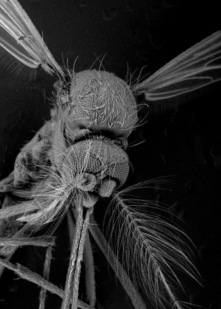
This Aedes aegypti mosquito came from the lab of Steve Whyard in biological sciences. To get this 200x magnification image, Huebner chilled the mosquito in a freezer so it remained still for imaging. This mosquito can spread diseases, like dengue fever. Do you notice the hairs on its wings?
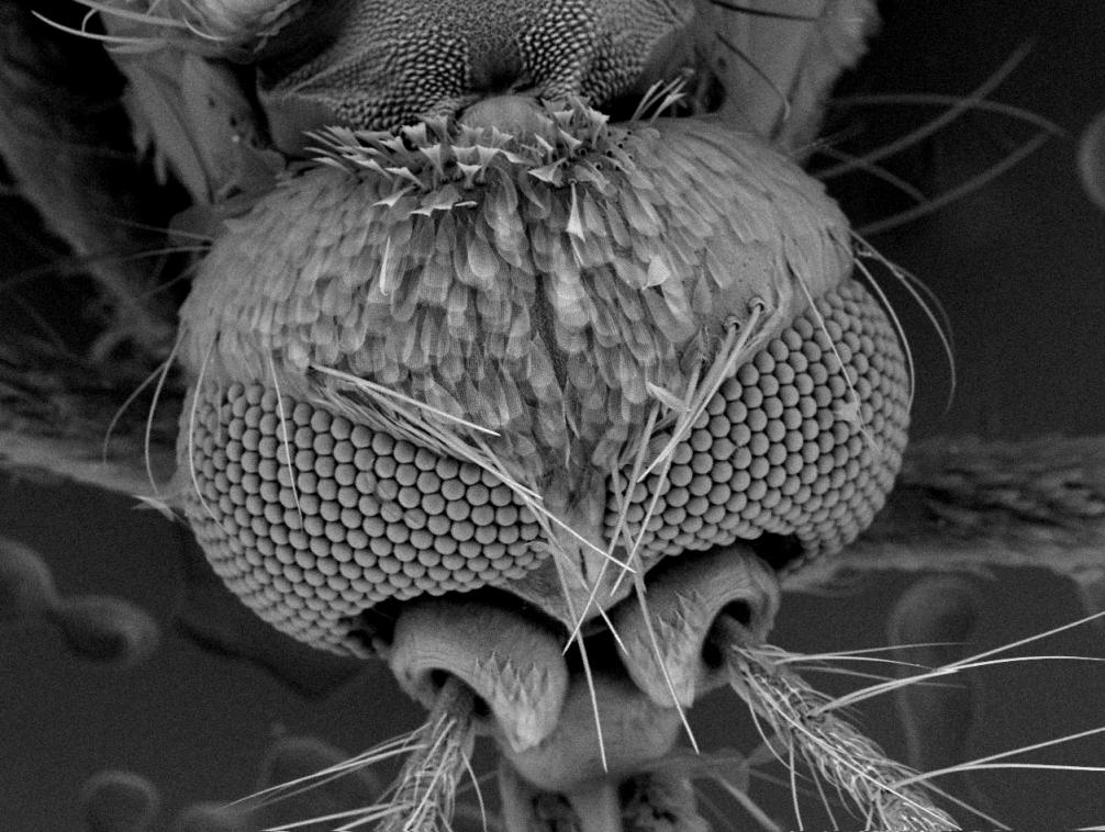
Scales above its eyes resemble plumage. The eyes — ommatidia — are found below, in a cluster of what seem like small ball bearings. Below those, you see the antennae coming out of their sockets.

A profile of the mosquito. The antennae, which look like Christmas trees, have remarkable abilities to pick up sound. A September 1999 paper in the Journal of Experimental Biology noted that the male of this species excels at picking up the vibrations made by a female flying. Indeed, the researchers said the sensitivity of the male antenna not only exceeds the sensitivity of the female antenna but also those of all other arthropod movement receivers studied so far. They also serve as chemoreceptors, detecting chemicals in the environment.
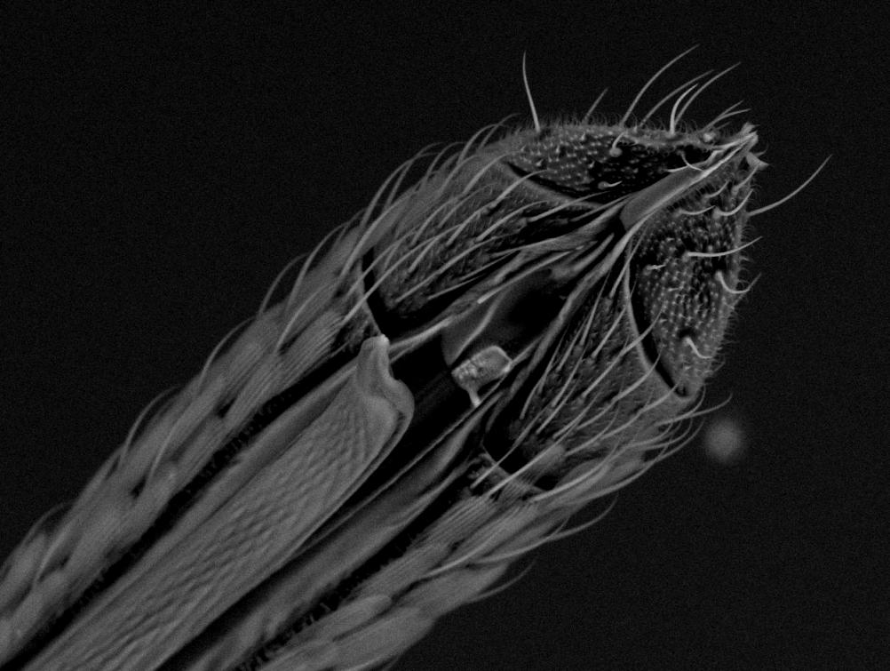
At 800x magnification, this is what a mosquito proboscis — the part that sucks your blood — looks like. The sheath with the hairs curls back and the needle extends to feed.
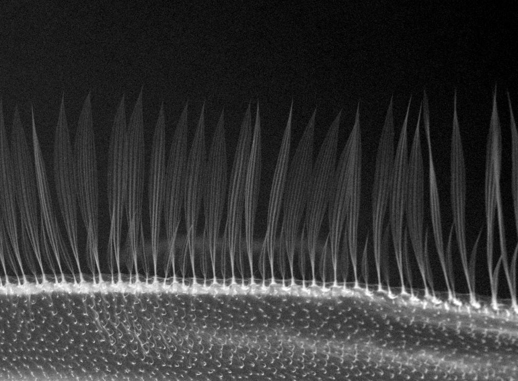
At roughly 1,000x magnification, one sees that on the back edge of this mosquito wing small hairs extend from it, perhaps serving some aerodynamic purpose.
Butterflies and moths
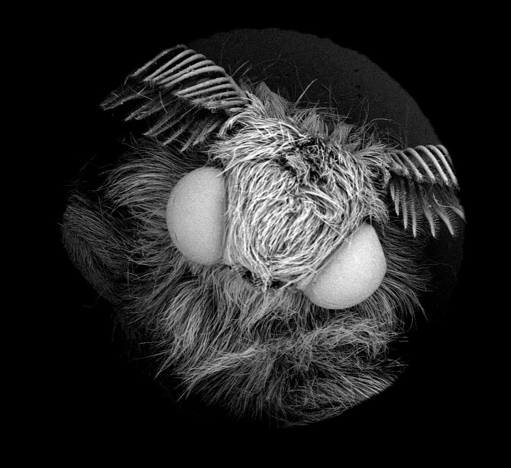
This is the face of a silkmoth. Moths outnumber butterflies and they are the ones who produce cocoons; butterflies make chrysalis.
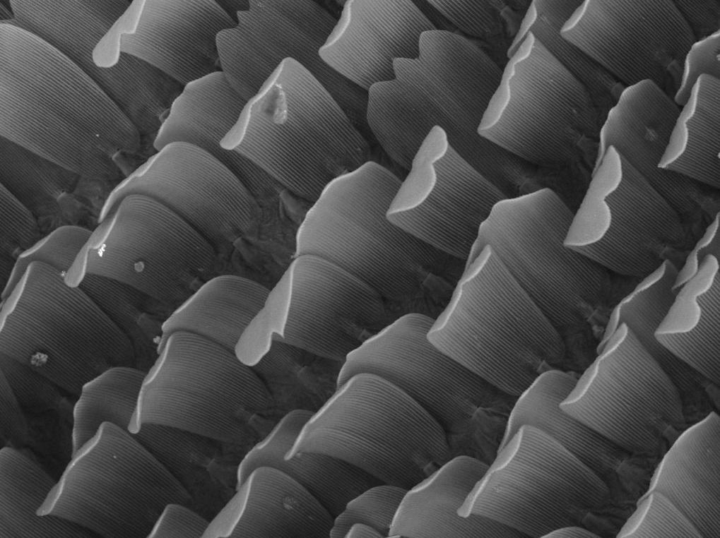
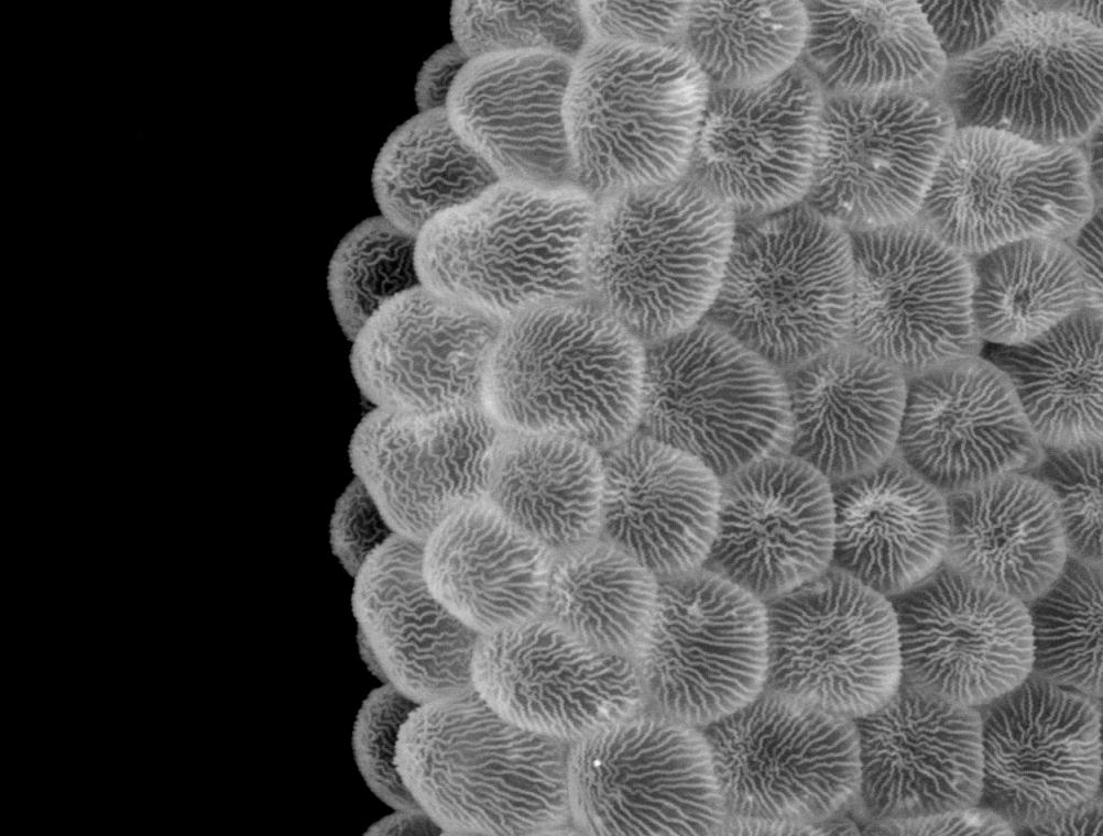
A common protein used in constructing insects is chitin. Here, one sees the arrangement of chitin on a butterfly wing. If you touch a butterfly wing (which UM Today does not endorse) and see a powder on your finger, it is this you have on your digits.
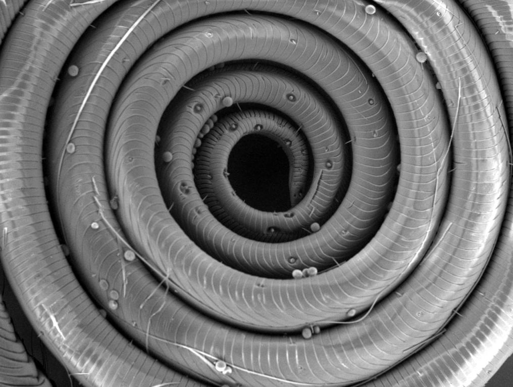
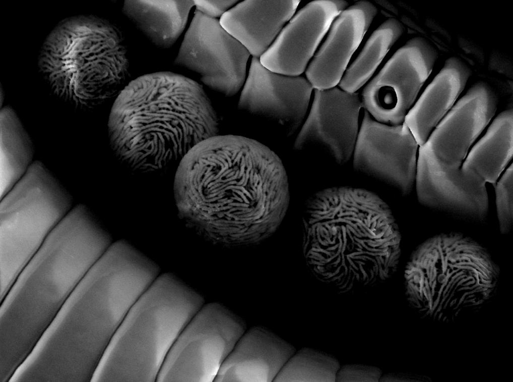
The coiled proboscis of a Hawk Moth. The small balls are pollen grains. The indented darkened spots are taste receptors.
Fly eye
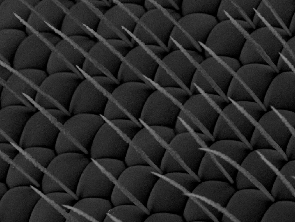
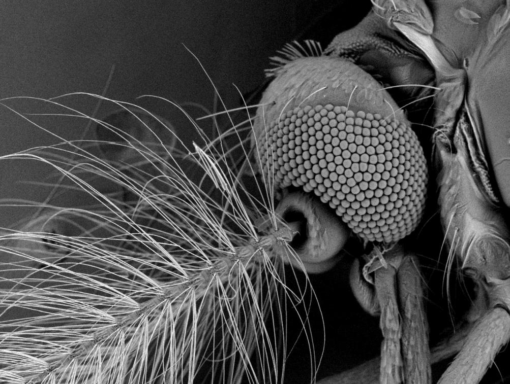
This is the eye of the Eristalis tenax, commonly called the Drone fly as they superficially look like drone bees. The fly has hairs protruding between individual ommatidium on its eyes. It’s uncertain what purpose they serve.
Shark skin
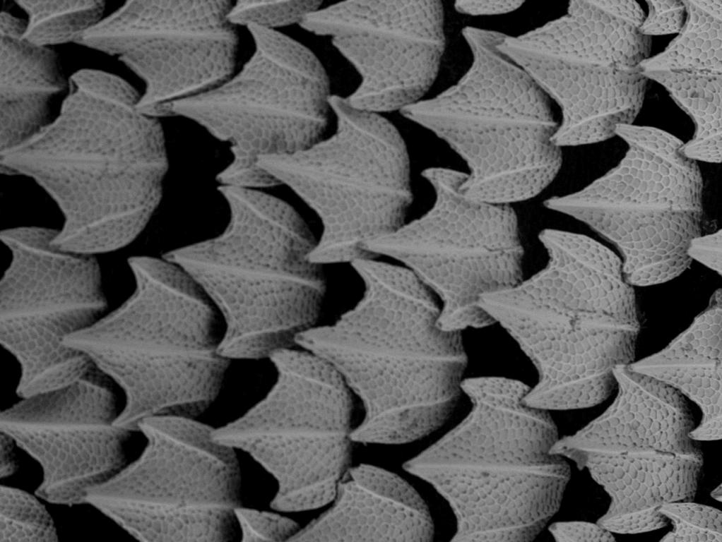
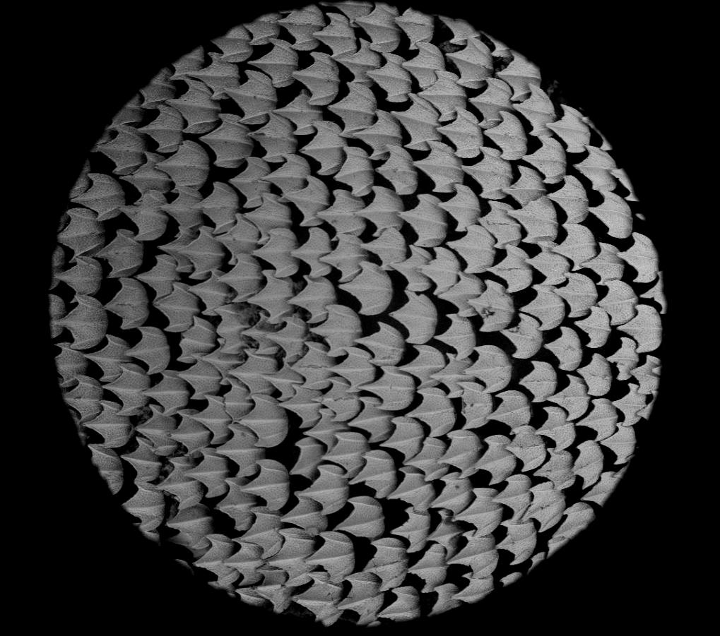
The placoid scales of shark skin, an ancient design. Stroke it one way, and it’s smooth; go against the grain, and it’s like sandpaper. And whereas many fish produce a mucous to help them slip through water, the placoid scales reduce hydrodynamic friction without needing this aid. Inspired by this, Speedo Inc. developed the Speedo Fastskin FSII swimsuit to reduce drag in water and counter the drag of human skin and hairs.
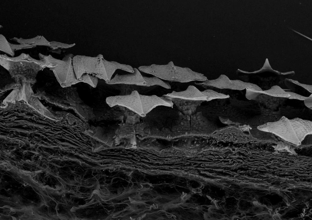
This cross section shows the underlying fibrous dermis and skin epidermis through which the placoid scales, which resemble unusually shaped teeth or birds in flight, protrude.
Plants
“As a biologist I have been privileged to be able to look at the microarchitecture of a variety of plants, fungi and animals and see a ‘world’ not visible to the naked eye by most who appreciate nature. The spectacular and wondrous structures that have evolved in nature have given us a rich biodiversity that surrounds us and allowed the myriad species to survive and flourish in many habitats. The overall intricacy and inherent beauty one sees in nature has inspired art and design for millennia.” — professor Erwin Huebner, writing for The Prairie Garden in 2013.
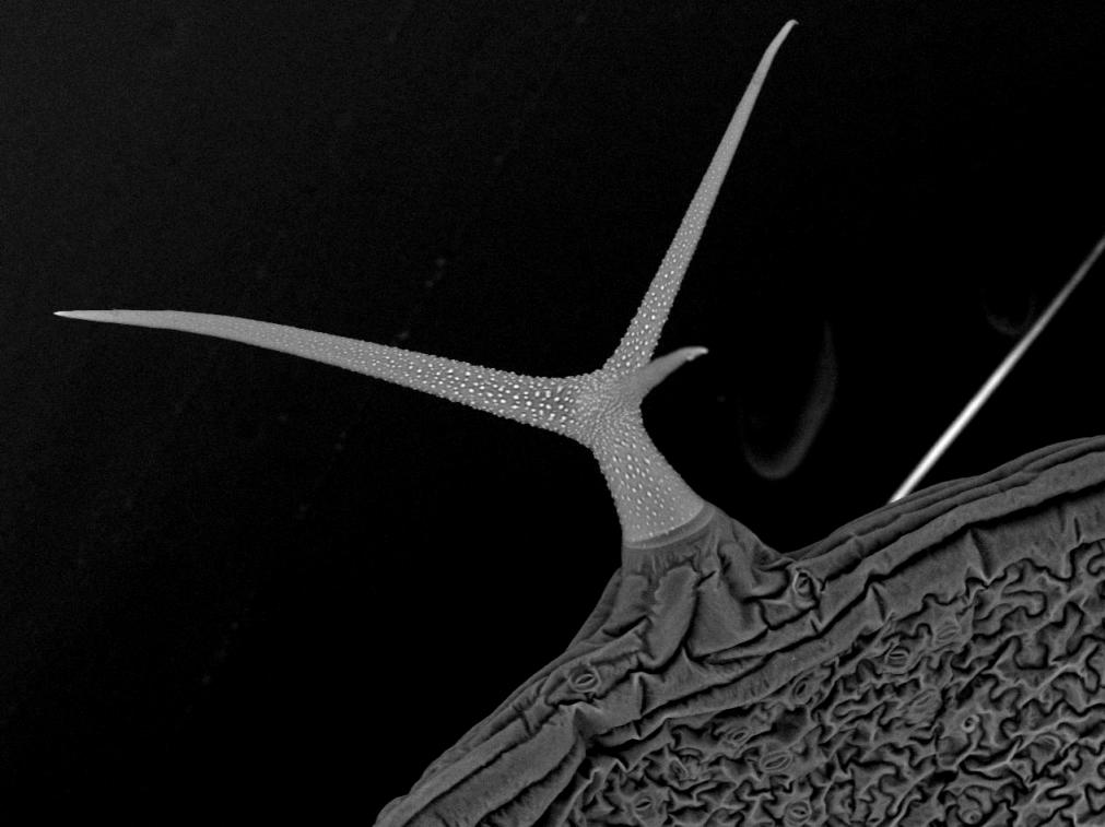
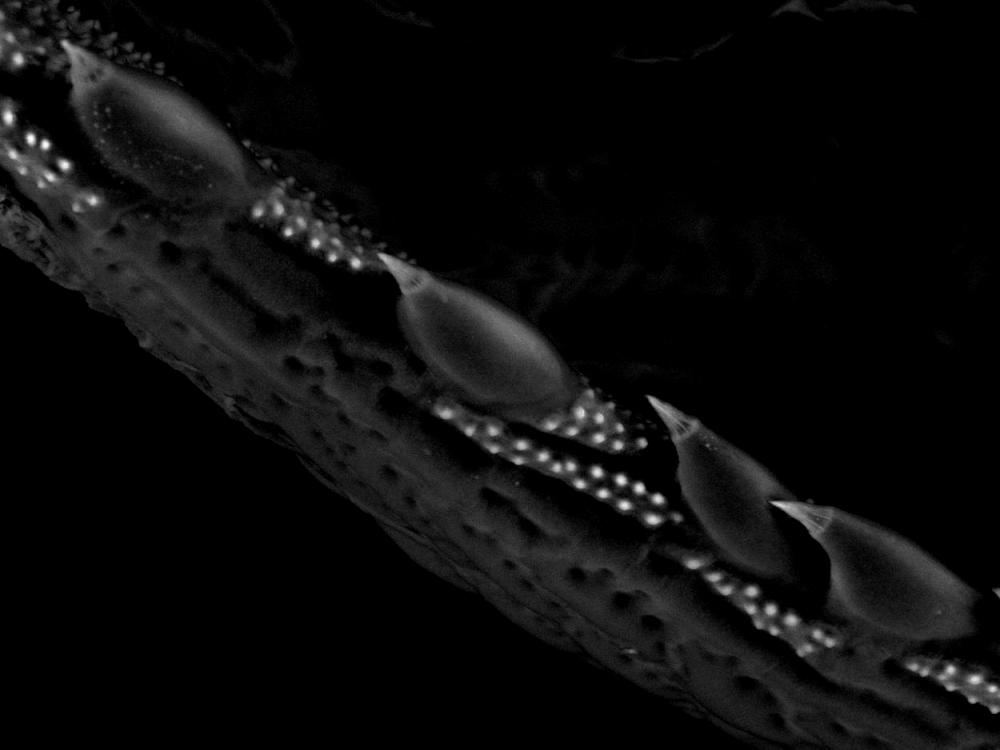
This is the Arapidopsis leaf’s trichrome. A trichrome is a hair on a plant leaf that often serves a defensive purpose, some can inject toxins (think of stinging nettle) but the Arapidopsis trichrome uses a different tactic to repel feeders. By its very structure, the trichrome offers mechanical protection, and It produce anti-feeding chemicals to deter insects that either try to eat part of the leaf or brush up against it.
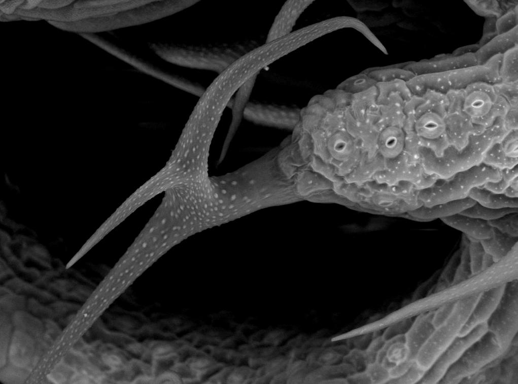
An Arapidopsis surface trichrome. The holes — stoma — on the right of the structure are used for gas exchange.
“Every aspect of Nature reveals a deep mystery and touches our sense of wonder and awe.”
— Carl Sagan, cosmologist
Error thrown
Object of class WP_Error could not be converted to string


