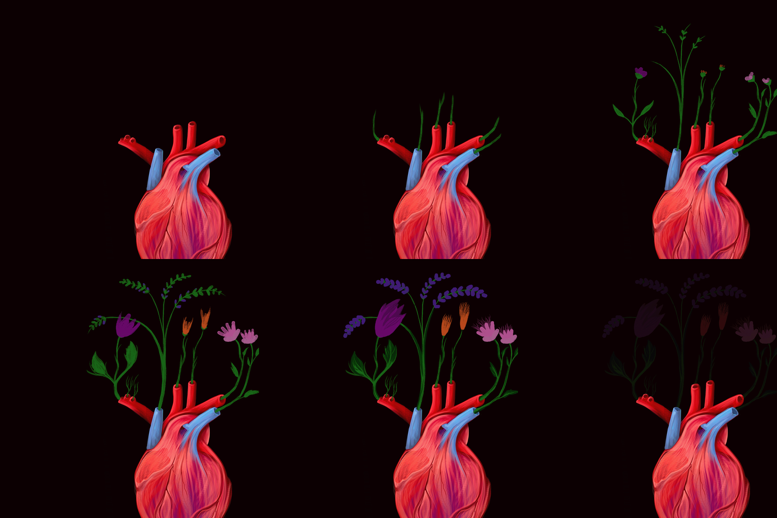
What Happens When We Donate our Body to Science?
We posed the question to Alexa Hryniuk, assistant professor in UM’s department of human anatomy and cell science.
First off, an acknowledgement that not everyone’s comfortable talking about cadavers. Hryniuk invites you to think of it another way.
“We refer to our body donors as ‘silent teachers,’” she says. “The students learn a great deal from the individuals who have graciously donated their bodies to our program.”
The initiative is a crucial element of the department, serving all five colleges at the Rady Faculty of Health Sciences. And the hands-on learning of human anatomy is an essential part of every medical student’s training.
Each year the department receives 30 to 40 donors. Bodies arrive within 24 hours after death and are quickly embalmed. Two techniques are employed: one which preserves the body for four years, which is the maximum time that the cadavers are kept on site, and a soft embalm where the body is kept only for four months, a technique designed to serve the specific needs of surgical residents.
In addition to first-year and graduate medical students, the cadavers are used by students in the dental program, the physician assistant program, the pharmacy program, as well as by physical therapy, midwifery and kinesiology students.
While students can participate in dissections, the majority are done by the head prosector—the official name of the individual tasked with preparing or dissecting anatomical subjects for demonstration—and teaching faculty. For example, a heart will be prepared for students to hold and manipulate so they can view the coronary arteries and all the valves and vessels inside the thoracic cavity, and observe how the heart interacts with the lungs.
“It really gives the students an appreciation of the three-dimensional space that anatomical structures take up that they can’t get anywhere else. Virtual reality programming is getting better, but it really doesn’t replace the touch and feel of these structures. The vast majority of our students will be doing hands-on surgical work with the human body, so to be able to feel and manipulate different portions of the body is a huge win for them,” notes Hryniuk.
As remarkable new forms of virtual technology continue to emerge some question if cadaveric dissection is still worth the time, money, and effort. “At the University of Manitoba we embrace both sides of how to teach anatomy. We have invested in the technology side as well. We find that it enhances learning,” explains Hryniuk.
However, she believes there are lessons gleaned from human dissection that can’t be replicated by tech, such as discovering that anatomical parts differ from person to person in terms of size and shape. Not everyone is the same, a distinction that is not clear from peering at textbooks and iPads.
Exposure to death and the emotion that arouses is another important lesson that students gain from working with a human cadaver. “It helps them reconcile that this will be part of their career and it also helps create a sense of empathy,” says Hryniuk.
Although students are expected to develop a sense of clinical detachment to help them to perform, staff also look for ways to develop empathy for their subjects. “When we find a patient with a pathology, that is a great teaching moment,” says Hryniuk. “We can not only talk about the pathology and how it may have come to be, but also discuss treatments the patient might have to go through and how to talk to the patient about the various pathologies they may have. It involves looking at the patient as a whole instead of individual parts or the ailments that they may have had.”
When the silent teachers reach the end of their service, they are cremated and honoured with a memorial service. Typically, students, teachers and the donor’s family attend. “Students will speak about their experiences in the anatomy lab and their experiences about learning from the donors. It is really quite beautiful how they describe how the donor has shaped their learning. It gives many families peace to hear how much their loved ones have influenced our health-care workers,” says Hryniuk.
Learn more about UM’s body donation program.
Alexa Hryniuk was among the panelists of a discussion about body donors and their role in health education, as part of UM Knowledge Exchange (previously called Café Scientifique).
Are you interested in hearing more from experts on a range of topics? We’re calling all alumni and community members to join us for the upcoming season of UM Knowledge Exchange—a hybrid event series, in-person and online, that brings the public together with researchers and experts. Hosted by the Office of the Vice-President (Research and International), the initiative is part of the Café Scientifique international event series and the UM Learning for Life Network.


